MD5: Characterization of kidney organoids as a model system to analyze bacterial colonization
|
|
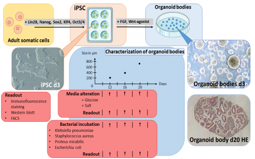
Organoids are three-dimensional cell cultures that resemble functional and structural traits of human organs. These structures have advantages over two-dimensional cell culture experiments given the establishment of complex polarized three dimensional cell-cell interactions that more closely mimic physiological barriers and functional processes in the human body. In our hands organoid structures develop following reprograming induced Pluripotent Stem Cells (iPSCs). Induced Pluripotent Stem Cells have been manipulated through retro- and lentiviral transduction of transcription factors such as Oct3/4, Sox2, Klf4, Lin28, Nanog and c-Myc causing them to regain their pluripotency when incubated with basic fibroblast growth factor (bFGF) and develop into kidney-like organoids following stimulation with a Wnt-agonist in 12 to 20 days. This project first aims to standardize the generation of kidney organoids. Kidney specific proteins and cell types are visualized and quantified by Western blotting, immunofluorescence staining and fluorescent activate single cell sorting. A comparison of different batches and different stages of organoid growth will be performed to assess variability in generating kidney organoids. High glucose and high salt addition to culture media and their influence on organoid generation are another focus of this project. Establishment of an infection model will be initiated after completion of standardization. For that means organoids of a chosen age will be incubated with bacteria such as Proteus mirabilis, Klebsiella pneumoniae, uropathogenic Escherichia coli tribes and Staphylococcus aureus, as the most common pathogens of acute infectious pyelonephritis. The bacterial colonization and infection will be monitored for bacterial numbers (multiplicity of infection) and spatiotemporal distribution throughout the organoid. Thus, novel insights into cellular responses following bacterial dissemination and cell damage as well as alterations in the barrier integrity will be gained. Furthermore, cell damage will be quantified by propidium iodine and annexin V staining as well as examination of damage and pathogen associated molecular patterns. Cooperation with other members of the RTG include Project 8, Prof. Dudeck’s, Prof. Naumann’s and Prof. Kahlfuß’ workgroup, as well as Prof. Kaasch and Prof. Zautner from the Institute of Microbiology.
Process of generating kidney organoids. After transforming adult somatic cells into iPSCs, these are held in culture on a basement membrane matrix. To set the conversion into kidney organoids in motion the iPSCs are stimulated for three days. Following that period, the organoid bodies will start to grow in size. On day 7 first tubular structures start appearing. Between day 12 and day 20 the differentiation of tubular segments reaches its peak and characterization as well as infection experiments are conducted in this time. From day 21 and onwards central fibrosis will arise due to deficient nutrient perfusion of the continuously growing organoid body. |
Photos: by UMMD, Melitta Schubert/Sarah Kossmann


